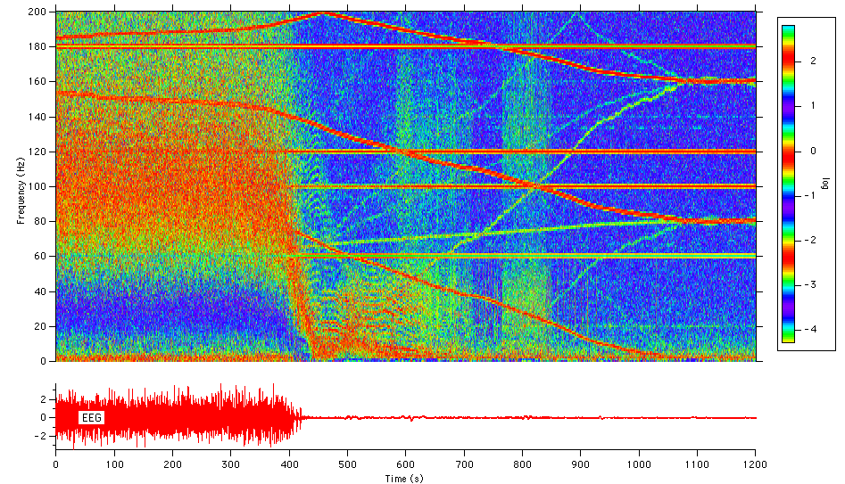

Cortical electroencephalogram (EEG) recorded in rat during anesthetic overdose. False-color power spectral image illustrates frequency distribution of the EEG, as well as presence of primary line noise and harmonics. Overdose initiated at 300 s.
Submitted by:
Dr. Roger Gallegos
Dept. of Neuropharmacology
The Scripps Research Institute

Forum

Support

Gallery
Igor Pro 9
Learn More
Igor XOP Toolkit
Learn More
Igor NIDAQ Tools MX
Learn More


